Multiscale alterations and cortex-wide organizational trends in focal cortical dysplasia

Multiscale alterations and cortex-wide organizational trends in focal cortical dysplasia
Sahlas, E.; Chen, J.; Dascal, A.; Levesque Kinder, B.; Favre, C.; Zhou, Y.; Royer, J.; Guberman, G. I.; Xie, K.; Lam, J.; Fadaie, F.; Pana, R.; Hall, J. A.; Ruber, T.; Weil, A. G.; Bernasconi, A.; Bernasconi, N.; Bernhardt, B. C.
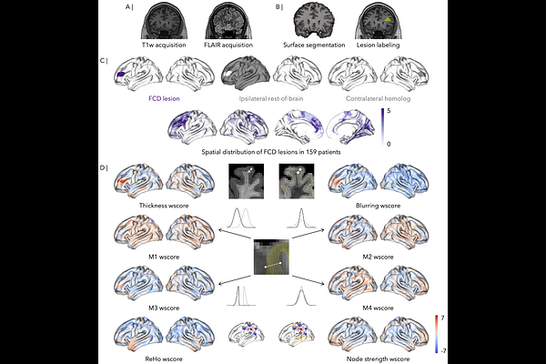
AbstractBACKGROUND AND OBJECTIVES. Focal cortical dysplasia (FCD) is a leading cause of surgically remediable pharmaco-resistant epilepsy. Previous research has used various MRI sequences to profile FCD lesions, but multi-site studies using personalized features derived from sequences routinely used in the clinic are sparse. This study aimed to quantify changes in cortical morphology, myeloarchitecture, and function within FCD lesions, compare MRI signatures between lesion subtypes and outcomes, and investigate links to macroscale brain organization. METHODS. This cross-sectional study included patients with FCD-related epilepsy aggregated across 3 datasets from Canada and Germany. The 3T MRI acquisitions included T1-weighted and fluid-attenuated inversion recovery (FLAIR) sequences, with the addition of a resting-state functional sequence for two datasets. We derived cortex-wide maps of patient-specific variations in morphology, myeloarchitecture, and function. Variations were quantified in lesions, ipsilateral cortex, and homotopic regions using age- and sex-adjusted normative models. Subgroup analyses explored the role of histological subtype and surgical outcome. Variations were also related to anterior-posterior and sensory-association organizational gradients. RESULTS. We included 159 patients with epilepsy and FCD (47 % female) and 183 healthy control participants (50 % female). Lesions exhibited changes in morphology (cortical thickness; FLAIR blurring) and in specific measures of depth-dependent intracortical myelin (variance and kurtosis of intracortical myelin profiles across depths), but not in local function (regional homogeneity; node strength). Spatial contextualization indicated more pronounced thickness changes in transmodal association cortices, while increased blurring of the gray-white matter interface co-localized with posterior cortical regions. FCD Type IIb lesions contained more marked changes in blurring and depth-dependent intracortical myelin (kurtosis of intracortical myelin profiles across depths) than Type IIa lesions. DISCUSSION. Our findings provide robust evidence that multiscale MRI profiling can identify FCD signatures and contribute to in-vivo subtyping. Furthermore, the novel use of myeloarchitecture profiling and contextualization with macro-scale brain gradients provides new avenues to understand intracortical alterations and the embedding of FCD lesions into broader organizational patterns.