Coupling between Notch signalling and junctional mechanics during asymmetric division of sensory organ precursors

Coupling between Notch signalling and junctional mechanics during asymmetric division of sensory organ precursors
PINOT, M.; Roland, L. B.
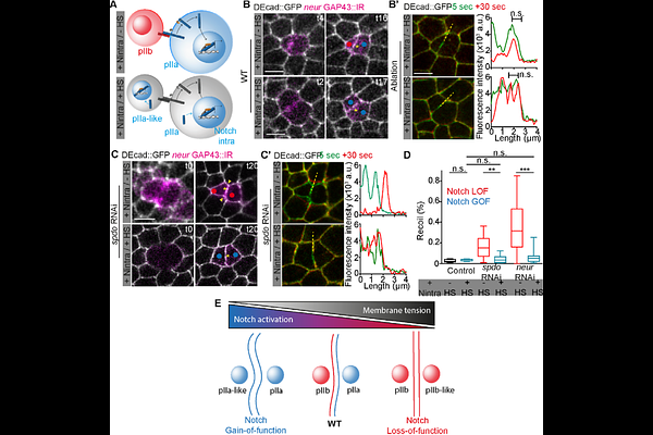
AbstractMechanical forces and signaling pathways are increasingly recognized as interdependent regulators of epithelial morphogenesis, yet their combined role in cell fate acquisition remains poorly understood. Here, we investigate the interplay between adherens junction mechanics and Notch receptor signaling during the asymmetric division of sensory organ precursors in the Drosophila pupal notum epithelium. Using quantitative live imaging and laser ablation, we identify the newly formed interface between SOP daughter cells as a mechanically specialized junction, characterized by persistently low membrane tension, distinct adhesive organization, and a unique cortical actomyosin architecture. We propose that low membrane tension may facilitate efficient Notch activation, as ligand-mediated endocytosis promotes Notch signaling by generating traction forces of a few piconewtons, oriented perpendicular to the plasma membrane. Perturbations of Notch pathway activity systematically alter junctional recoil following laser ablation, with reduced Notch signaling correlating with increased tension. Conversely, constitutive Notch activation in a Notch loss-of-function context is sufficient to restore a low-tension state. These findings suggest that Notch signaling actively shapes the mechanical properties of its signaling interface, indicating reciprocal interactions between mechanics and signaling. Together, our results support a model in which Notch activity and junctional mechanics are coupled during asymmetric cell division, highlighting how local mechanical states may contribute to the robustness of cell fate specification in epithelia.